YOUR MORPHOLOGY CELL ASSISTANT
YMCA. Built around the expert.
An AI-assisted workflow for peripheral blood smear review, with a focus on abnormal or uncertain cells, from candidate detection to expert confirmation.
THE WORKFLOW
STEP 01
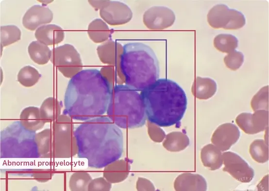
Locate and segment candidate cells.
The research pipeline detects candidates within the supplied image, records their positions and prepares cell images through cropping and segmentation. Failed or uncertain outputs require review.

STEP 02
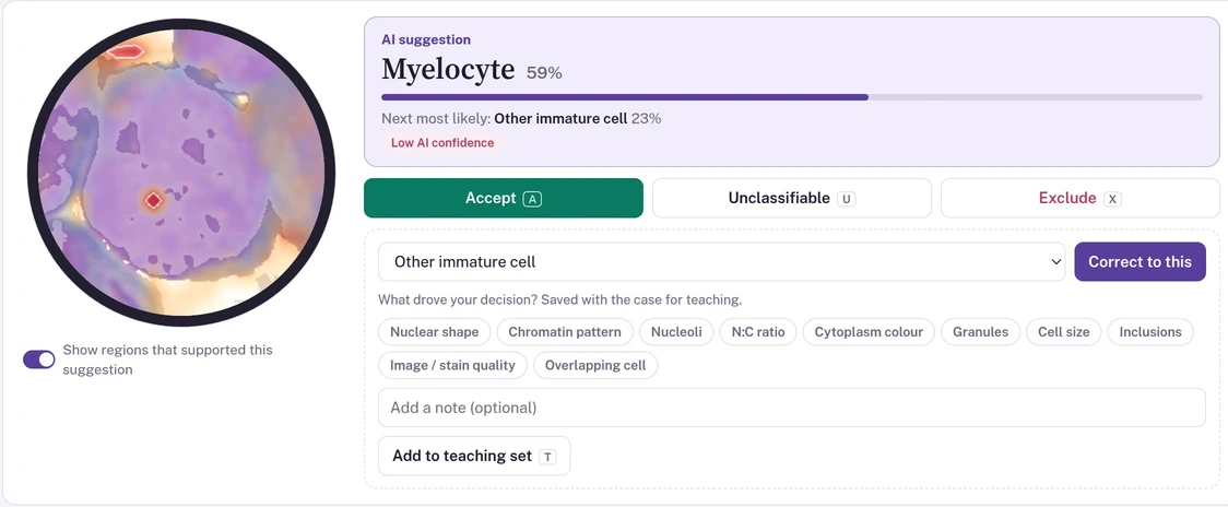
A shared starting point for review.
The classifier offers 16-category morphology pre-classification, including mature and immature cell types, labels suggestive of abnormal morphology, and artifacts. Model confidence and quality flags guide expert review; category labels alone do not establish a normal or abnormal finding, or a diagnosis.
STEP 03
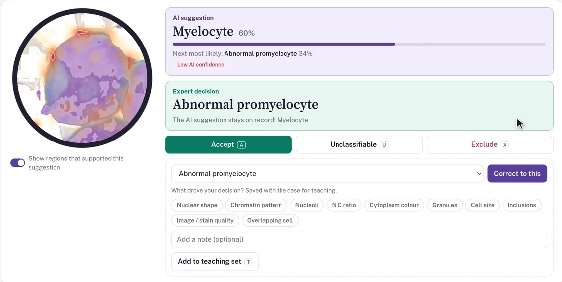
Correct a label. Recover a missed cell.
Professionals can confirm, relabel or exclude a candidate. A missed candidate can be marked on the original image for a subsequent analysis rerun.
STEP 04
Keep the prediction and the correction.
AI outputs and human review events are saved separately. The prototype aggregates classification counts and review status into research summaries.
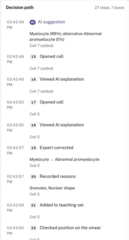
Image analysis covers the supplied image area. It does not guarantee that the entire physical slide was acquired or that no cell was missed.
ASSISTANCE THAT STAYS INSPECTABLE
Context for review decisions.
The prototype presents the original image location, segmentation output, predicted class, model confidence scores and quality flags together. Professionals can return to the source image and inspect what the model analyzed.
AI assistant for review
The research assistant answers questions about a case or cell in plain language, such as which cells need review, or which morphology information and reference cells relate to an AI suggestion. Its answers are generated from the cell information and reference-cell comparisons shown on screen and are automatically checked for consistency with them; this is not expert or clinical validation. Review changes the assistant proposes are applied only after a professional confirms them. These aids are not validated diagnostic evidence; professionals remain responsible for final interpretation.
Digital review history
The original model label stays separate from the human correction. Each review event records the previous and updated review state, with notes and timestamps for retrospective analysis.
Quality flags for review
Quality flags highlight uncertain predictions and potential image-analysis issues, helping professionals identify results that need closer inspection. Results with uncertainty flags cannot be accepted in batch.
SEE YMCA IN ACTION
A closer look at the prototype.
Product walkthrough from the team’s research prototype, with audio and English subtitles. Shows image analysis, morphology explanations, reference-cell comparisons, assistant answers with action confirmation and recorded expert review actions.
PRODUCT DEVELOPMENT
Designed for expert review. Clinical validation ahead.
Today, the local prototype accepts static image files and supports session-based cell review and research report drafts. The updated walkthrough also shows review replay and expert-led teaching cases. Planned product capabilities include formal patient and specimen identity, role-based access, clinical sign-off, equipment integration and LIS/HIS exchange.
Explore the validation plan